Punstoppable
A list of puns related to "Thoracic Aorta"
Edit: After meeting with the doctors I am going through with both operations simultaneously. They seem very confident. They have done it together 6 times previously with no difficulty and I will be the 7th. The surgery was scheduled for 10am this morning but there was an emergency that required Roselli to stay up all night and needed to sleep to preform my operation fresh. So it is rescheduled for tomorrow at the same time. My nerves are shot after all f that. This has been an insanely difficult rollercoaster of events. Thank you everyone.
I'm 39 and aortic aneurysms run in my family (dad's side). I'm on pain meds but will do my best to answer!
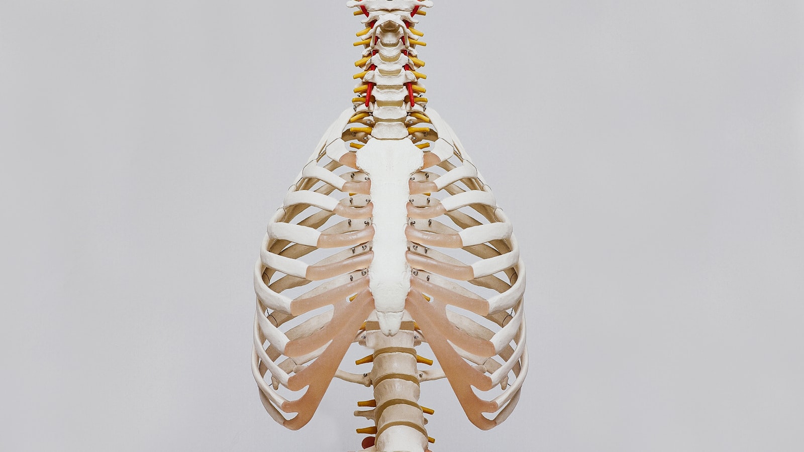
Hi, I’ve read a little on how the aorta influences the development of scoliosis but am still having trouble understanding it. The descending aorta curves down on the left side of the thorax, around the level of t3/t4. It runs left lateral to the vertebral bodies at the midthoracic level (t4-t8) and is positioned more anteriorly to the vertebral bodies from t9-l2. A 2003 study showed that in right thoracic idiopathic scoliosis, the aorta is positioned more laterally and posteriorly relative to the vertebral body, and the distance from the posterior aspect of the aorta to the anterior aspect of the spinal canal was less in the scoliosis group as well. So, with thoracic scoliosis being overwhelmingly more common on the right side and the aorta lateralized to the left of the thorax, this begs the question of what its exact role is in the development of curves. The first thing that comes to mind for me is the possible asymmetry in blood flow. Although the aorta has a relatively low surface area, it branches out from all sides during its descent down the spine. This coupled with the fact that many spinal muscles originate from the more dorsal bony projections of the vertebrae themselves could result in one side of the spinal muscles becoming more metabolically active than the other. The only problem I see here is that the left-sided aorta should reasonably facilitate the left spinal muscles, but in right thoracic scoliosis we see the opposite pattern(?). Right thoracic scoliosis is usually associated with right hand dominance and stronger, facilitated thoracic spinal muscles on the right side. So this is where my reasoning hits a wall and the concept of scoliosis becomes even more confusing. Can anyone offer any insights? My own case becomes even a bit more confusing. Born right dominant, developed left thoracic in teens and somewhere along the way became stronger and more dominant with my left hand/arm. Could this imply more aortic involvement in left thoracic cases?
F 55 yrs, chest X-ray report said, among other things,
"There is an artherosclerotic aorta and degenerative changes of the thoracic spine."
The doctor said the spine thing was arthritis but walked off when I asked about the aorta. I've tried Google but the results are so mixed I don't understand them. Some Google results talk about an abdomen aneurism and I'm confused. I'm 7 yrs older than my dad's age when he died of a heart attack w hardening of the arteries, in case that's related somehow.
What is it, and what should I do, please?
Thank you in advance!
I am a Surgical Technologist in a Hybrid operating room. I just found out that on Monday, I will be scrubbing a case that has never been done at my facility. Here's the info I have:
Patient will be on bypass. Cardiothoracic surgeon will be performing whole case, and as of right now there is not a vascular surgeon scheduled to assist. The open heart team will get the pt cannulated, then they will perform an Aortic Root Repair. At that point, I will be scrubbing in to assist with the insertion of a thoracic aorta stent. The plan is not to access femorally, but to do a retrograde stick.
This is where I am unclear. where will the stick be, brachial? Or since the chest will be open, are there other options? Has anyone done/seen a similar case? I would like to know what to expect, as of last night, the doc was not forthcoming with more details of his plan. I really hate not being prepared, and I don't want to "wing it" on a case that sounds so complicated.
The pt has a dissection all the way from thoracic aorta to the iliacs, and I am unsure if he plans to stent anywhere below the thoracic aorta.
Thanks for any insights.
Age / 30 Sex / M Height / 5'6 Weight /168 Race / White Duration of complaint / About a year Location (Geographic and on body) live in Florida / interior of chest Any existing relevant medical issues / none known prior to this diagnosis. Current medications / Xanax .5 for what my primary care believed to be anxiety.
No family history due to me being adopted.
I am a Police Officer of 10 years in a very violent area. I am under constant stress from the call volume and violent nature of alot of calls I go to. I have been training for about a year to prepare myself for SWAT team try outs. It is the most intense training I have ever done in my life. I have lost almost 30 pounds. Under exercise I become extremely short of breath especially with circuit style training. I will usually vomit during training, and have an exceptional thirst. I chalked it up to possible asthma. I began to experience severe jaw pain after exercise it felt like my jaw was locking, then it began to effect the back of my neck. This happened several times. I then experienced a panic attack. The first in my life. I went to my primary care and was prescribed lexapro and xanax. I took the lexapro for about a month but it made me feel "out of focus" so I stopped taking it. The Xanax was prescribed for emergency panic attacks or sleep, as I was experiencing insomnia that my primary care thought was due to the anxiety. I still have the xanax but do not take it unless I cannot sleep in the middle of the night. that occurs less than one time every few weeks.
I had an EKG done at my primary and it came back abnormal but did not point to anything specific, I was referred to a Cardiologist. The cardiologist has a terrible bedside manner. I've seen him 3 times and our visits have lasted for less than 10 minutes in total (seriously I'm not exaggerating). He was annoyed at me not knowing my family history. He ordered a EKG, ECHO, and a Cardiolite stress test. Those tests were done and I visited again yesterday and was told everything was fine except I had Ascending Thoracic Aorta dilation of 4.2cm he told me not worry about it until it got to 5cm. He then told me to not lift weights anymore and to have a low sodium diet, then left the room. I literally chased him down the hallway asking questions about what it meant, I did not feel like it was explained to me where I could understand it. He told me that he was ordering a CT to confirm the diagnosis and that I was to come back with the results. I ha
... keep reading on reddit ➡Just finish speaking with my thoracic surgeon and it’s time. After so many years of waiting to see when I’ll have this surgery, it’s now happening. My aorta is measuring 4.9. I only blame myself for this. After finding out my condition, I still lived life as if I was normal. I couldn’t accept my body. I hated it. Now I only wished I would have treated it with love and care, and kept a calm and peaceful life.
Find peace within yourself my friends.
So, I've got this old fic on AO3 which was really more a collection of wild theories and headcanons about the Owl House, and I thought it'd be cool to share some of the ones related to the Bile Sac and how magic could be a biological function. Assuming that it's not just a gland that secretes magic juice.
Based on the diagram Eda showed Luz, it looks like the bile sac is connected to the left atrium, which receives the oxygen rich blood from the lungs and pumps it to the rest of the body via the aorta.
Furthermore, assuming that the diagram is anatomically correct, it would mean that the bile sac takes up quite a bit of space inside the thoracic cavity. Since most animals have fairly limited space when it comes to their internal organs, and most witches don’t have disproportionately large torsos, it’s possible that either have smaller lungs or just one.
Since there's magic everywhere in the Demon Realm, it’s safe to assume that the plants and wildlife are saturated with the stuff.
https://www.mdpi.com/1420-3049/26/19/5914/htm
Some highlights in no particular order:
> "Among the substances investigated, the essential oils of Myristica fragrans (nutmeg), Heracleum transcaucasicum, Heracleum anisactis, Anethum graveolens (dill), Apium nodiflorum, Petroselinum crispum (parsley), Pycnocycla bashagardiana and Piper sarmentosum, all containing high concentrations of myristicin, ranging between 12% and 96% of the composition, are noteworthy. "
>
>" An interesting publication discusses the anticonvulsant and inhibitory effects on glial activation of Myristica fragrans (nutmeg) extract. This material, containing about 11% myristicin, was tested in male NMRI rats that were induced to have seizures. Behavioral studies have shown that pretreatment with nutmeg extract effectively reduced seizure behavior, decreased cell death in the hypothalamus and improved glial activation [71]. "
>
>" A publication on the aqueous extract of the aerial part of parsley (Petroselinum crispum) sought to investigate the antihypertensive activity of the plant. In vivo studies were performed with male albino rats, and an in vitro study used isolated thoracic aorta rings. The results show a potent vasorelaxant activity in aortic vascular rings, while in animals the extract induced a decrease in blood pressure parameters. More detailed studies showed that there was a blockage of calcium channels present in the vascular wall, but also suggest that other pathways may be involved in the antihypertensive effect such as, for example, increased nitric oxide synthesis [67].
>
>The ability of myristicin to protect neurons from hypoxia-induced injuries was investigated. To conduct these assays, rat dorsal root ganglion (DRG) neurons were used. The results showed that myristicin reduced the viability of neurons when exposed to concentrations greater than 50 mM. However, at lower concentrations, it significantly increased cell viability in neurons when exposed to hypoxia, as it protected against hypoxic injury, not causing apoptosis. Complementary trials showed that myristicin decreased cleaved caspase-3 and bcl-2 levels in these hypoxia-induced neurons. Therefore, it was observed that it can reverse hypoxia-induced apopto
39/F 5’2” 240lbs Non-smoker Don’t drink No recreational drugs
Current Diagnosis’s: GERD and IBS since 1997 Anxiety and Depression since 2007 Raynauds since 2010 Hypothyroidism since 2014 Plantar fasciitis since 2017 Migraines/Atypical Facial Pain since 2018 Mild Carpal Tunnel since 2019 Allergies to environments/foods so I immunotherapy since 2019 COVID in Dec 2020 and Dec 2021
Current Meds: Levothyroxine Protonix Claritin Flonase Singulair RA Allergy Plaquenil Vitamin D CoQ10 Prenatal vitamin
Family History: Mom- Lung cancer, COPD, Emphysema, Diabetes, High blood pressure, and Hypothyroidism. Dad died at 38 from unknown causes, but Heart Disease runs on his side of the family.
I’ve been on the hunt for what’s wrong with me for several years. The symptoms keep piling on and every Dr looks at me like I’m crazy. Here are the various symptoms I’ve had over the past couple of years:
Since forever Frequent urination, especially at night
1997 Acid reflux
2008 Heart palpitations
2010 Raynauds Mystery bruises Sciatica on right side
2012 Ankle and leg swelling
2015 More Mystery Bruises Weight gain got bad (100lbs from 2015-2020) Plantar fasciitis began
2017 Finger swelling Random hives Burning Right side Flank Pain
2018 Finger nail beds dark bands (Terry’s Nails), receding (Onycholysis), and splinter hemorrhage marks Migraines/Atypical Facial Pain Hand/finger numbness began Shortness of breath when taking stairs or exercising began Achy/tight upper back Split-Second Fainting Sensations C-Reactive Protein (CRP) chronically elevated from 6-14
2019 Writer’s cramps pain Finger joint pain
2020 Clenching teeth began Shortness of breath continued -can’t walk and talk at the same time -tightness in lower back and ribs Got COVID
2021 Trip to ER in April -sudden double vision and dizziness -trouble swallowing -sudden tiredness (hard to keep my eyes open) -cat scan of brain clear Achilles’ tendon pain began July Headaches/Migraines resumed in Aug Hand/Finger numbness resumed in Aug Achy lower back began in August Achy/tight upper back resumed in Sept Dry mouth and eyes began Sept Sinus pressure/swelling/stuffy Dry cough began Nov 5th Shortness of breath got worse beginning in November Unexplained weight loss Got COVID again Increased heart rate Right eye pain and blurrier than normal vision
————————————————————
Saw 3 Rheumatologists since 2018 -one said I have RA, but other 2 disagree -ran all blood tests that came back normal except ANA IFA 1:80
... keep reading on reddit ➡Hey all. To preface the medical history, I had heart surgery in 2009 on a VSD. Have had no problems since.
I’ve been trying to enlist in the Marines since August of 2020, got sent to MEPS in September, scored very very good on the PiCat, made IST qualifications, made it through the medical physical until the doctor told me I would need to have my paperwork sent to BUMED (despite the doctor having approved my paperwork). The reasoning on the BUMED waiver that my recruiter has shown me is: Thoracic Surgery, Bicuspid Aorta, Aortic Insufficiency, and unspecified RBBB. The first waiver was shot down after weeks of waiting and constant hassle of MEPS from my recruiter trying to hear back. I was extremely devastated but wasn’t ready to quit so I called my doctors office to get more information and some words from my cardiologist, who had already given me a clearance letter saying, verbatim, that he “sponsors my enlistment into the Marines, and that I’m asymptomatic from a cardiovascular standpoint”. The doctor said he wasn’t able to do much more, but the office sent me my last visits notes. In it were some more details, and more intimate notes from the doctor. I sent it in and was again denied.
My problem is this: the disapproval letter says nothing behind the reasoning, it simply says the request for a medical waiver is disapproved. Reading other posts online, I had figured they usually gave reasoning? I was curious if anyone could provide insight on how to get in touch with BUMED to plead my case/get clarity on the grounds for disapproval, or any other alternatives, maybe like a MEPS doctor consulting me? I’ve considered a congressional inquiry, but otherwise I feel very hopeless.
Sorry for the long post, thanks to all.
37F , stopped smoking 2.5 years ago, not currently taking any medications. Scan done after a small nodule found on my lung while having a different test on lower abdomen. Won't see my doc for another month to discuss results, just curious what this means basically. Thanks for any help you can provide!
EXAM: DIC CT CHEST W/O IV CONTRAST
CLINICAL INDICATION: R91.1 (Solitary pulmonary nodule) Z87.891 (Personal history of nicotine dependence)
TECHNIQUE: Multiple axial images were reconstructed in 1 and 3 mm intervals from the thoracic inlet through the upper abdomen. Coronal and sagittal reconstructed images as well as stacked axial MIP images were generated from this data set. Dose reduction protocols were utilized
COMPARISON: None
FINDINGS: There is no evidence of axillary, supraclavicular, or mediastinal adenopathy. There is no significant pericardial effusion. The aorta is normal in caliber. Heart size is within normal limits. There is no significant pericardial effusion.
Limited imaging of the noncontrasted upper abdomen is unremarkable. No acute abnormality is seen. There is no aggressive osseous lesion. There is no evidence of malalignment in the spine.
There is a tiny 2 mm nodule in the right upper lobe on series 3 image 63. There is a well-circumscribed nodule abutting the minor fissure on the right on series 3 image 124 measuring 7.3 x 4.0 mm. There is a tiny nodule just anterior to the previous nodule on series 3 image 124 measuring 2.6 x 2.2 mm. There is a pleural-based nodule in the right middle lobe on series 3 image 151 measuring 6.3 x 5.9 mm. There is an opacified airway in the right middle lobe on series 3 image 184. There is a pleural-based nodule in the posterior sulcus on the right on series 3 image 265 measuring 5.0 x 4.1 mm. This corresponds to the nodule seen on the prior CT abdomen and pelvis. This nodule is stable. There is a calcified granuloma in the left upper lobe on series 3 image 117. There is a noncalcified nodule in the lingula on series 3 image 174 measuring 3.6 x 2.9 mm. No other nodules are seen.
Type 2 diabetes mellitus is the main risk factor for cardiovascular diseases. It has been reported that the reduction of mitochondrial protein sirtuin protein 3 contributes to the development of type 2 diabetes mellitus by impacting mitochondrial respiration.
Cordycepin is an adenosine derivative and is isolated from the culture filtrate of Cordyceps Militaris. This study explored the protective effect of cordycepin on vascular impairment induced by type 2 diabetes mellitus and its property mechanism.
In this study, a type 2 diabetes mellitus rat model was established. The endothelium-dependent relaxation of the thoracic aorta ring decreased in type 2 diabetes mellitus rats that could be reversed by cordycepin. Next, the mitochondrion impairment in human umbilical vein endothelial was detected by JC-1 staining.
The in vitro studies reveal that cordycepin plays a beneficial role in advanced glycation end products-induced endothelial mitochondrion impairment. Moreover, according to the cordycepin molecular docking analysis, cordycepin can bind to sirtuin protein 3. Cordycepin increased the expression and activation of sirtuin protein 3 in a dose-dependent manner. sirtuin protein 3 interruption blocked the protective effect of cordycepin on mitochondrion in human umbilical vein endothelial.
Cordycepin can conclusively protect vascular function impaired by type 2 diabetes mellitus, and the mechanism may potentially be involved in the sirtuin protein 3 signal pathway.
From :
- https://www.dl.begellhouse.com/references/708ae68d64b17c52,forthcoming,41927.html
I had orchiectomy in March, 3xBEP in April, RPLND in September. I see oncologist Monday for first post scan meeting. Last few weeks my remaining testicle has felt somewhat heavy, sore. It hasn't grown to my knowledge but I'll ask him for an ultrasound. Can cancer reappear that quickly into other testicle?
My post RPLND scan and xray:
A 0.5 cm nodule within the posterior lateral left lower lobe is not
seen on the March 2021 examination. This could be
infectious/inflammatory, but close attention to this area on
follow-up imaging is needed. A tiny subpleural nodule within the
anterior inferior right middle lobe is unchanged. Several small areas
of airspace consolidation are present within the posterolateral right
lower lobe. A few subtle patchy groundglass opacities are seen
superior to this site. This is favored to represent an
infectious/inflammatory process. No pleural effusion is present. The
central airways are patent.
Heart size is normal. There is no pericardial effusion. The thoracic
aorta is normal in caliber. The tip of a right chest port catheter is
at the superior cavoatrial junction. No pathologically enlarged
mediastinal, hilar, or axillary lymph nodes are identified. The
thyroid gland appears unremarkable.
Abdomen/Pelvis:
The liver enhances homogeneously. There is no biliary ductal
dilatation. The gallbladder is nondilated. The spleen, pancreas, and
left adrenal gland appear unremarkable. There is a 1.4 x 1.1 cm right
adrenal nodule which is unchanged from prior examinations.
Enhancement of the kidneys is symmetric. The abdominal aorta is
normal in caliber. Multiple surgical clips are visualized within the
retroperitoneum status post resection of the left retroperitoneal
mass and retroperitoneal lymph node sampling. No new pathologically
enlarged retroperitoneal or mesenteric lymph nodes are identified.
There is a small fat-containing periumbilical hernia.
The bowel is normal in caliber, without focal wall thickening or
evidence of obstruction. The appendix is nondilated. No free
intraperitoneal air is identified. The rectum and urinary bladder
appear unremarkable. The prostate gland measures near the upper
limits of normal. There is no free pelvic fluid. No pathologically
enlarged pelvic lymph nodes are identified.
Age 22
Weight: 190lb
Race: Caucasian
Height: 6ft 2 inches
Time of complaint: 3 weeks
Medication none
Biological Sex: Male
Here is the copy paste of the whole report:
Study Date11/16/2021 Technical quality: Good visualization Type of Study: TTE procedure: M-Mode, Doppler , Color Doppler, Adult Trans Thoracic Echo 2D Complete, Myocardial Strain. Priority:RoutineHR: 59 bpmBP: 150/78 mmHg Conclusions Summary Transthoracic real-time echocardiogram, complete, with 2-D and m-mode recording, pulsed and continuous wave spectral doppler, color flow velocity mapping, and myocardial strain performed, with the following findings:
Findings Mitral Valve The mitral valve is normal. There is no evidence of mitral stenosis or regurgitation. Aortic Valve The aortic valve is trileaflet with normal structure. There is no evidence of aortic stenosis or regurgitation. Tricuspid Valve The tricuspid valve is normal. There is no evidence of tricuspid stenosis or regurgitation. Insufficient TR jet to estimated PASP. Pulmonic Valve The pulmonic valve structure appears normal. There is no evidence of pulmonic stenosis or regurgitation. Left Atrium Normal left atrial volume index. Left Ventricle The left ventricle is normal in size, wall thickness, wall motion, and systolic function. Calculated LVEF is 59%. Average global longitudinal strain (GLS) -16%. Normal LV diastolic function. Right Atrium The right atrium size is normal. Right Ventricle Normal right ventricular cavity size and systolic function. Pericardial Effusion No evidence of pericardial effusion. Miscellaneous Aortic root and proximal ascending aorta are normal in size. Atrial septum not well visualized. Normal IVC size with normal respirophasic collapse. Aortic arch appears normal. Valves Mitral Valve Peak E-Wave: 0.6
... keep reading on reddit ➡39 male 5’4” 145lbs
I have an thoracic aortic dilation at the small aneurysmal stage. I am given to understand that the only suggested activity modifications relate to intrathoracic pressure, shear pressure, and arterial blood pressure.
I think the valsalva maneuver causes a spike in intrathoracic pressure for some reasons that I never figured out. Does a reverse simple valsalva cause some drop or do something to the heart that’d strain the aorta? I’m talking about closing the epiglottis and inhaling hard, sucking hard through a straw, or forceful inhalation playing a wind instrument like the harmonica.
Any thoughts please?
Wife 39yo is diagnosed stage 3, possible stage 4. HR+, HER2-, 10cm tumor, couple other additional 2cm tumors and some swollen lateral lymph nodes up to her collar bone, whole left side is as advanced as stage 3 can be. Two concerning very small nodes that DR's are unsure are mets or inflammation but suggest that it might be mets based on PET and CT scans. One is in the back on a rib and the other near the aorta in mid chest and unable to biopsy either. Second opinion with a top radiologist believes early stage 4 but also not clear.
Our current situation is:
A. Treat as stage 3 aggressively. But the oncologists said outcomes and life span aren't as good if it's actually stage 4 and you burn your body out with all the aggressive treatments up front since it'll just spread again.
B. Treat as stage 4 starting with hormone blockers. Only using chemo as needed and rotating through medicines over the next few years. Hope that a stage 4 trial comes a long that applies.
C. Treat as stage 3 with ispy2 trial (or similar national trial) with standard chemo + AC, radiation, surgery route but a bonus breast cancer drug that is shown to work well in other cancer settings and matched to her cancer subtype.
D. Wait 3-8 weeks for more biopsy results, chest node surgery and results, genetics to all come back then decide a,b,c. The road we're currently on.
We're waiting to speak with a cardio thoracic team to use a robot to biopsy the chest node, which is in a dangerous spot. There's also an ICD involved that needs to be removed which adds a couple more week wait after surgery. Suggesting Lupron while we wait.
My wife and I are of the opinion that she's a strong fighter and young enough that the hormones and wait stage 4 route is dumb. That we should start chemo immediately and go after it aggressively and see what happens. We feel much more pressure timeline wise if we're on the cusp for 3 to 4. We have an amazing marriage a 1yo and a 4yo and this is fucking it all up. It's like the worse dream we could conjure up. We don't want to not be aggressive only to find out that oops it spread more in a few months / years after it didn't response to a drug only to wish we went hard out of the gate.
Anyone else have similar predicaments? What were your deciding factors, thoughts, outcomes and experience with the road you chose?
Hi. I was in a car accident today and had to get a chest CT done. After I got home from the hospital, I was reading my CT report. My doctor didn’t mention anything about it, so i can’t imagine it’s a concern but I’m curious as to what it means. I am obese, so im not sure if that has any significance.
This is the reading in question “MEDIASTINUM/AXILLAE: Heart size is normal without pericardial effusion. The thoracic aorta is normal in caliber. There is anterior mediastinal triangular soft tissue density with interspersed fat consistent with residual thymic tissue.”
The last sentence is what had me a bit baffled and not understanding what it means. If anyone could explain that to me, it would be great.
Before anyone tells me to ask my doctor, I don’t see him for awhile to follow up (he’s booked), and I’m genuinely curious and want to learn. I’m not asking for advice, just want to learn terminology. I’m going to discuss everything with my doctor when I see him.
Thanks in advanced.
3/31/21. Original Post
A couple of days ago, I awoke to find my daughter in an awake, eyes open, and enlarged, but mute, unresponsive state. SOne eye seemed bigger than the other or set forward more somehow.She had repetitive, stereotyped movements, like bracing herself on her arms, pushing herself upward repeatedly, to no effect. Her mouth was open with a fish-like look to it. Panic, fear, surprise in her eyes/face. Her arms were alternately folding up to the chest, fists rolling inward, and then curling into the lap, wrists curled inward. Her legs were extending and rolling inwards. I'm not sure she was breathing. Then that all stopped and she just curled over and didn't move. We called 911.
At the hospital:
History: "Patient with a possible episode of seizure and following altered mental status. "
Her oxygen level was 77%; She didn't have a gag reflex, one pupil was 3mm, one was 4mm.
| CBC Day of event 10:22AM | Shortly after | Discharge |
|---|---|---|
| WBC 21.5 ** | 27** | 6 (after IVIG, +steriods) |
| RDW 12.3 | 12 | 12.1 |
| RBC 4.07 | 3.78** | 3.75** |
| PLT 693**** | 540**** | 479**** |
| MPV 8.4** | 8.3** | 8.3** |
| MCV 94.3 | 89.7 | 93.6 |
| MCHC 32 (then 31.6**) | 31.6** | 33.3 |
| MCH 30.2 | 29.5 | 29.9 |
| HGB 12.3 | 11.5** | 11.3** |
| HCT 38.4 | 33.9** | 33.9** |
BiBasilaer Alterlisis
"Minimal subpleural consolidation in the dependent portions of the lower lobes likely related to atelectasis with aspiration possible given secretions within the trachea and mainstem bronchi."
Phosphorus 7.2,
10:22 EGFR 85 mL/min, Lipase 31,
| VBG 10:22AM | 10:22 | 11:42 Intubation |
|---|---|---|
| PH 7.3** | 7.11 ** | |
| PCO2 52 mmHg** | 84 ** | |
| PO2 73 mmHg** | 59 ** | |
| Sodium 136 mmol/L, | 137 | |
| Potassium 4.5 mmol | 4.0 | |
| Chloride 103 mmol | 103 | |
| Calcium++ 1.24 mmol/L | 1.34 ** | |
| Glucose 140 mg/dL** | 160 ** ? why | |
| Lactate 0.7 mmol/L | 1.1 | |
| HgB 11.5 g/d** | 12.4 | |
| HCO3 26 mmol/L | 27 | |
| ANION GAP 7 mmolL | 7 mmol/L | |
11:06 AM VBG's PH 7.3***, PCO2 52 mmHg***, *PO2 73 mmHg*******,, Sodium 136 mmol/L, Potassium 4.5 mmol, Chloride 103 mmol, Calcium++ 1.24 mmol/L, Glucose 140 mg/dL** Lactate .7 mmol/L, HgB 11.5 g/dL** , HCO3 26 mmol, Anion Gap 7 mmol/L
Anion Gap 11 (then 13**, 16**) Calcium 9.7 Chloride 104 CO2 25 (then 22, 19) Creatinine 0.94. (then 1.05**) Glucose 153. Po
... keep reading on reddit ➡Do your worst!
For context I'm a Refuse Driver (Garbage man) & today I was on food waste. After I'd tipped I was checking the wagon for any defects when I spotted a lone pea balanced on the lifts.
I said "hey look, an escaPEA"
No one near me but it didn't half make me laugh for a good hour or so!
Edit: I can't believe how much this has blown up. Thank you everyone I've had a blast reading through the replies 😂
It really does, I swear!
Hi, I’ve read a little on how the aorta influences the development of scoliosis but am still having trouble understanding it. The descending aorta curves down on the left side of the thorax, around the level of t3/t4. It runs left lateral to the vertebral bodies at the midthoracic level (t4-t8) and is positioned more anteriorly to the vertebral bodies from t9-l2. A 2003 study showed that in right thoracic idiopathic scoliosis, the aorta is positioned more laterally and posteriorly relative to the vertebral body, and the distance from the posterior aspect of the aorta to the anterior aspect of the spinal canal was less in the scoliosis group as well. So, with thoracic scoliosis being overwhelmingly more common on the right side and the aorta lateralized to the left of the thorax, this begs the question of what its exact role is in the development of curves. The first thing that comes to mind for me is the possible asymmetry in blood flow. Although the aorta has a relatively low surface area, it branches out from all sides during its descent down the spine. This coupled with the fact that many spinal muscles originate from the more dorsal bony projections of the vertebrae themselves could result in one side of the spinal muscles becoming more metabolically active than the other. The only problem I see here is that the left-sided aorta should reasonably facilitate the left spinal muscles, but in right thoracic scoliosis we see the opposite pattern(?). Right thoracic scoliosis is usually associated with right hand dominance and stronger, facilitated thoracic spinal muscles on the right side. So this is where my logic hits a wall and the concept of scoliosis becomes even more enigmatic. Can anyone offer any insights? My own case is a bit puzzling. Born right dominant, developed left thoracic in teens and somewhere along the way became stronger and more dominant with my left hand/arm.
Please note that this site uses cookies to personalise content and adverts, to provide social media features, and to analyse web traffic. Click here for more information.