
Punstoppable
A list of puns related to "Fluorescence Microscopy"
I get fluorescence idea where something with a lot of conjugations absorbs light at longer wavelength and then emits light at lower wavelength. Why aren't the absorbance and emission same? Where does energy stay after being absorbed that accounts for difference with emission
https://link.springer.com/article/10.1007/s00775-015-1306-y
The main purpose of the following study was the determination of elemental changes occurring within hippocampal formation as a result of high-fat and carbohydrate-restricted ketogenic diet (KD). To realize it, X-ray fluorescence microscopy was applied for topographic and quantitative analysis of P, S, K, Ca, Fe, Cu, Zn and Se in hippocampal formations taken from rats fed with two different KDs and naïve controls. The detailed comparisons were done for sectors 1 and 3 of the Ammon’s, the dentate gyrus and hilus of dentate gyrus. The results of elemental analysis showed that the KDs induced statistically significant changes in the accumulation of P, K, Ca, Zn and Se in particular areas of hippocampal formation and these alterations strongly depended on the composition of the diets. Much greater influence on the hippocampal areal densities of examined elements was found for the KD which was characterized by a lower content of carbohydrates, higher content of fats and increased proportion of unsaturated fatty acids. The levels of P, K and Zn decreased whilst those of Ca and Se increased as a result of the treatment with the KDs.
Hello! I am a Graduate student at Arizona State University. I am trying to develop a small kit that people can receive in the mail to have their cheek cells imaged using cool fluorescence microscopy. Its purely for fun and the novelty. I always thought the cheek cell lab from high school bio was unsatisfying when you see other beautiful examples of microscopy, so I developed this little kit so people can have their cheek cells imaged in their full glory. My background is in molecular biology and microscopy, so I do all the imaging myself. Here are some examples of what I have been able to get. Again this is just for fun and gives you no info on your health.
blue nuclei green cytoplasm 100x
green nuclei blue cytoplasm 200x
blue nuclei red cytoplasm 400x
If anyone is interested in helping me test out this kit it would be much appreciated. I am not charging anything right now. I am just looking for people to let me mail them the kit and try it, so I can see if it can survive shipping without any changes in quality. You can choose your own two colors!
TLDR: let me send you a free kit to image your cheek cells with fluorescence microscopy. I need people to try it out so I can work out the kinks
Edit 7/8/21: Thanks to anyone who showed interest in this post! I appreciate all the help, getting something like this off the ground is really hard, but I have successfully gathered volunteers! for now I wont need anymore, but I will update this post later once I have the kits available to the public
Hi! This might be silly question from my side, but I cannot understand one concept. So, as I know, fluorescence absorbs light and then emits it. I am wondering, when fluorescence is used in microscopy, how can you track molecules or observe some changes that has duration of for example milliseconds, if fluorescence can be lost in nanoseconds ? Do you use multi photon fluorescence to excite at consequent time frames, or how does it work? This have been on my mind quite some time now and I would really appreciate any explanation from you
I am reading papers where confocal fluorescence microscopy images were analysed. In many of the papers I see the term "puncta" being used when researchers analyse the colocalisation between 2 proteins. For example, in the linked paper:
>In addition, many C1q-positive puncta in the IPL were associated with synaptic puncta identified by double immunostaining with synaptic markers such as PSD-95
I am not sure what is meant by the term "puncta" in the context of immunocytochemistry. I have read that the general definition for puncta is a small, distinct point. However, I was wondering whether puncta is a synonym for clusters of a protein (e.g. in the example above, does synaptic puncta refer to clusters of the PSD-95 protein?)
Any insights are appreciated.
Journal of the American Chemical SocietyDOI: 10.1021/jacs.1c03642
Yi Zhang, Haonan Zong, Cheng Zong, Yuying Tan, Meng Zhang, Yuewei Zhan, and Ji-Xin Cheng
https://ift.tt/3z59bnS
Consider an inverted epi-fluorescence setup imaging fluorophores in water through a glass coverslip using a high NA oil immersion objective.
I have always assumed that the effectively used NA of a microscope system is limited by the lowest refractive index medium between the sample and the objective lens. This of course includes the mounting medium, and an NA1.4 oil immersion objective will actually only give NA1.33 for samples suspended in water (still talking about my assumption).
But then it struck me that other effects may come into play when fluorophores are very close to the substrate surface. For, e.g., labeled RNA immobilized to the surface, the fluorophores may sit a few tens (maybe hundreds) nm from the glass surface. Would near field effects like evanescent coupling or something else give higher angle wavefronts in the substrate than what’s predicted by Snell’s law? And hence allowing for higher effective NA than what’s possible for samples deeper in the mounting medium? If this is the case, at which distance from the surface would these effects come into play?
TLDR: What’s the highest obtainable NA for an oil immersion microscope lens when viewing fluorophores in water very close to the glass substrate surface?
Hierarchical porosity within a zeolite crystal is visualized by X‐ray scattering microscopy to probe nanoporosity within the inorganic zeolite phase. In complement, in situ fluorescence microscopy allows to monitor pore hierarchization in real‐time via organic fluorescent stains, which fill the meso‐ and macropore space.
Introducing hierarchical porosity to zeolites is vital for providing molecular access to microporous domains. Yet, the dynamics of meso‐ and macropore formation has remained elusive and pore space ill‐characterized by a lack of (in situ) microscopic tools sensitive to nanoporosity. Here, we probe hierarchical porosity formation within a zeolite ZSM‐5 crystal in real‐time by in situ fluorescence microscopy during desilication. In addition, we introduce small‐angle X‐ray scattering microscopy as novel characterization tool to map intracrystal meso‐ and macropore properties. It is shown that hierarchical porosity formation initiates at the crystal surface and propagates to the crystal core via a pore front with decreasing rate. Also, hierarchical porosity only establishes in specific (segments of) subunits which constitute ZSM‐5. Such space‐dependent meso‐ and macroporosity implies local discrepancies in diffusion, performance and deactivation behaviors even within a zeolite crystal.
https://ift.tt/3cHxIWf
Hi, I am doing immunocytochemistry experiments where I have done indirect immunofluorescence staining of fixed CHO cells against some proteins of interest. I have taken images of the cells using a confocal laser scanning microscope. I have been analysing my images using ImageJ.
In my analysis, I am outlining the cells of interest using the Polygon tool and then measuring the mean intensity of the proteins of interest in grayscale mode, so I can see any difference between the treated and control cells. I have done this experiment before and I am now doing a repeat of this.
However for this experiment, the density of cells on my coverslips is high and they are overlapping with each other & densely packed together. When I outline a cell of interest, I find that regions of it overlap with neighbouring cells. I have been told by some people that I can still analyse these cells (i.e. measure the mean intensity of my protein of interest) even if they do overlap with neighbouring cells due to the fact that the cells were grown as a monolayer and that I am not analysing any membrane proteins. They also said that it's ok if the cells overlap with parts of neighbouring cells so long as they don't significantly overlap with other cells (e.g. cell of interest overlaps with 50% of a neighbouring cell).
I am wondering if anyone else who has done immunocytochemistry experiments have encountered this kind of problem with overlapping cells and whether they think that it is ok to analyse the cells given this issue? Any insights are appreciated.
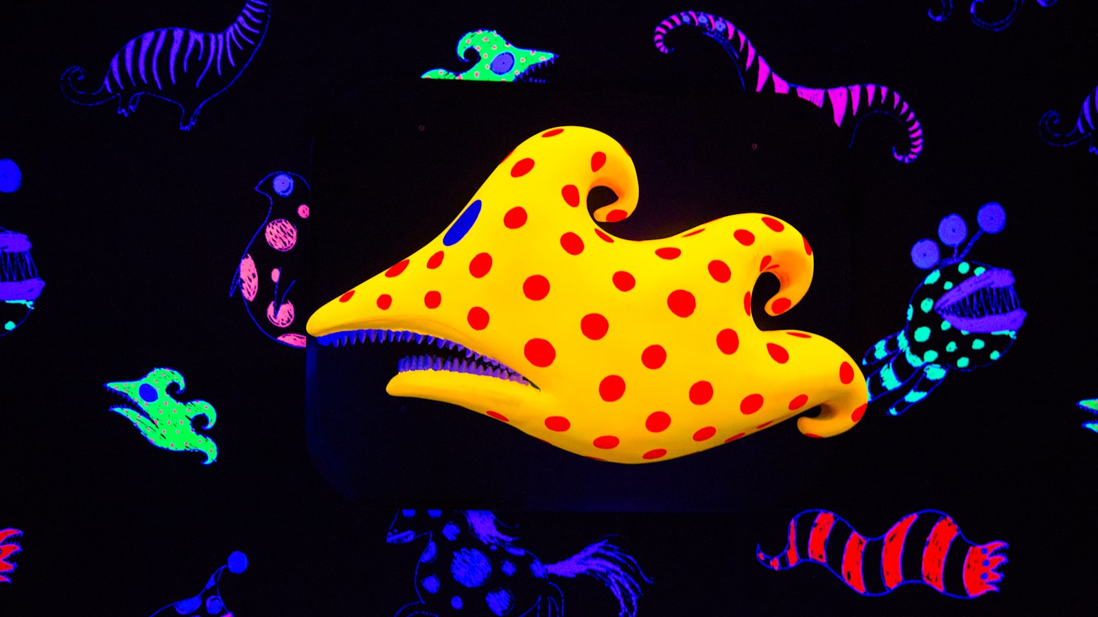
Zeolite catalysts often display highly intergrown structures, yielding a zeolite “flower” or “sun” at the microscale. Using small‐angle X‐ray scattering and in‐situ fluorescence microscopy, Bert M. Weckhuysen et al. show in their Communication (DOI: 10.1002/anie.202101747) that porosity is developed only in specific segments of zeolites, which might cause strong local discrepancies in performance and deactivation behavior. The yellow sun in black background is reminiscent of the symbol of Utrecht University, where the work is performed.
https://ift.tt/3h46QDW
Journal of the American Chemical SocietyDOI: 10.1021/jacs.1c00014
Tom Baladi, Jesper R. Nilsson, Audrey Gallud, Emanuele Celauro, Cécile Gasse, Fabienne Levi-Acobas, Ivo Sarac, Marcel R. Hollenstein, Anders Dahlén, Elin K. Esbjörner, and L. Marcus Wilhelmsson
https://ift.tt/3cIYdMg
This is my first time teaching this workshop and I honestly don't know how many slides I'll need for several sections with a total of 70 students, but I'm sure they'll drop quite a few. I don't have my own lab at this university to make the slides myself, so I'll have to buy them, but they're so incredibly expensive
P.S. I'm really sorry for spamming this sub, but I don't have anyone else to ask
Please note that this site uses cookies to personalise content and adverts, to provide social media features, and to analyse web traffic. Click here for more information.


